

 Nanomedicine
Journal of Extracellular Vesicles | In situ tumor vesicles carrying oncolytic adenovirus achieve long-lasting immunotherapy
QQ Academic Group: 1092348845
Detailed

 The study was published in Journal of Extracellular Vesicles under the title “In Situ Sustained Delivery of Tumor Cell-Derived Extracellular Nanovesicles With Oncolytic Adenoviruses for Potentiating Cancer Immunotherapy”.
Oncolytic adenoviruses (OVs) can directly eliminate cancer cells and subsequently trigger immune responses, exhibiting great potential for antitumor therapy. Nevertheless, it has been observed that immune cells may also undergo lysis during viral treatment, which markedly blunts OV-mediated antitumor immunity. In this work, we developed a microneedle (MN)-based platform with in situ generated tumor cell-derived extracellular nanovesicle (TDEV)-coated OVs to boost cancer immunotherapy and mitigate immune cell exhaustion. For this platform, tumor cells pre-infected with OVs are loaded into the upper reservoir of the microneedle device. After transdermal administration, hollow microneedles facilitate the continuous transport of in situ formed TDEV-coated OVs toward the tumor site for sustained OV delivery. These OVs selectively infect cancer cells rather than immune cells. Improved antigens generated from OV-induced tumor cell killing can be presented by non-exhausted dendritic cells, which further elicits robust immunotherapeutic effects in TC-1-hCD46 xenograft tumor-bearing mice and mouse models of postoperative tumor recurrence.
Quick Look at the Literature
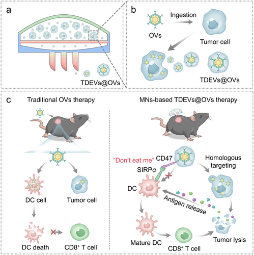
Figure 1
Microneedle-based in situ TDEVs@OVs platform for enhanced tumor immunotherapy and prevention of immune cell exhaustion. (a) Pre-infected tumor cells are loaded into the upper reservoir of hollow microneedles to achieve sustained in situ release of TDEVs@OVs. (b) Schematic illustration showing that tumor cells take up oncolytic viruses and continuously secrete TDEVs@OVs. (c) Schematic of microneedle-guided in situ secretion of TDEVs@OVs to potentiate tumor immunotherapy and restrain immune cell exhaustion.
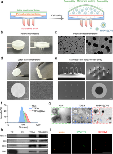
Figure 2
Fabrication and characterization of microneedles. (a) Schematic illustration of microneedle fabrication. (b) Representative photograph of microneedles. (c) Representative photograph of polycarbonate membrane; scale bars: 10 μm (left), 2 μm (right). (d) Representative photograph of latex elastic membrane; scale bars: 5 mm (left), 20 μm (right). (e) Representative scanning electron microscopy image of the microneedle array (stainless steel hollow needles); scale bars: 500 μm (top and bottom left), 50 μm (bottom right). (f) Particle size distribution profiles of exosomal vesicles, tumor-derived extracellular vesicles, and tumor-derived extracellular vesicles loaded with exosomal vesicles. (g) Transmission electron microscopy images of exosomal vesicles, tumor-derived extracellular vesicles, and tumor-derived extracellular vesicles loaded with exosomal vesicles. Scale bar: 100 nm. Red arrows: tumor-derived extracellular vesicles loaded with exosomal vesicles; blue arrows: tumor-derived extracellular vesicles. (h) Western blot analysis for the expression of TSG101, CD9, CD81 (tumor-derived extracellular vesicle markers), and CD47 (“don’t eat me” signal marker). (i) Colocalization observation of tumor-derived extracellular vesicles loaded with exosomal vesicles under laser scanning confocal microscopy. Exosomal vesicles were labeled with fluorescein isothiocyanate (green), and tumor-derived extracellular vesicles were labeled with CD9-Cy5 (red); scale bar: 100 μm.
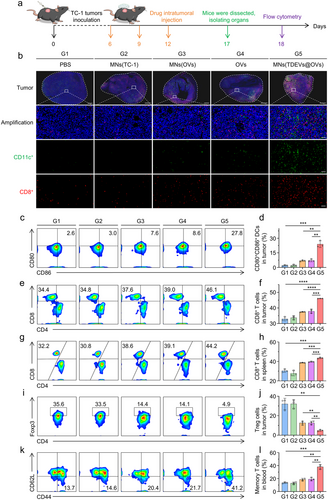
Figure 3
In vivo immune-activating capacity of microneedle-based in situ tumor-derived extracellular vesicle-wrapped oncolytic viruses. (a) Schematic illustration of the microneedle immunological evaluation experiment using female C57BL/6 mice bearing TC-1-hCD46 xenograft tumors. (b) Immunofluorescence images of CD11c-positive dendritic cells and CD8-positive T cells in tumor tissues. Scale bars: 1 mm (whole tumor view), 50 μm (magnified region). (c, d) Representative flow cytometry dot plots and relative quantification of intratumoral dendritic cells (CD45⁺CD11c⁺CD80⁺CD86⁺). (e, f) Representative flow cytometry dot plots and relative quantification of intratumoral CD8⁺ T cells (CD45⁺CD3⁺CD8⁺). (g, h) Representative flow cytometry dot plots and relative quantification of splenic CD8⁺ T cells (CD45⁺CD3⁺CD8⁺). (i, j) Representative flow cytometry dot plots and relative quantification of intratumoral regulatory T cells (CD45⁺CD3⁺CD4⁺FOXP3⁺). (k, l) Representative flow cytometry dot plots and relative quantification of effector memory T cells (CD45⁺CD3⁺CD8⁺CD62L⁻CD44⁺) in peripheral blood. Unpaired t-test was used for statistical analysis. Data are presented as mean ± SD (n = 3). P values: *P < 0.05, **P < 0.01, ***P < 0.001, ****P < 0.0001.
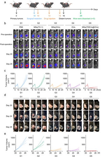
Figure 4
In vivo evaluation of microneedle-based in situ tumor-derived extracellular vesicle-wrapped oncolytic viruses for suppressing tumor recurrence and metastasis. (a) Schematic of the study on the inhibition of tumor recurrence and metastasis using microneedles (TDEVs@OVs) in female C57BL/6 mice bearing TC-1-hCD46 xenograft tumors. (b) Bioluminescence images of primary tumor resection and postoperative recurrence. (c) Individual growth curves of primary tumors in each group (n = 5). (d) Representative photographs of distant tumor metastasis. (e) Individual growth curves of distant metastatic tumors in each group (n = 5).
Reference Information
Journal of Extracellular Vesicles (IF 21.7)
Pub Date: 2026-01-06
DOI: 10.1002/jev2.70222
We specialize in custom nano-medical functional materials. We conduct targeted development to tailor structure, particle size and surface modification for your experiments. Full characterization, iterative optimization and technical support are available for targeted delivery, bioimaging and nanomedicine studies. Send your evaluation plans to us.
Engineer Zhang: 17715390137
Engineer Chen: 18914047343
- Previous: Journal of Controlled
- Next: Frontiers of Medical E


