Diabetes mellitus (DM) is a ubiquitous chronic disease characterized by persistent high blood sugar levels. Among diabetic patients, it is estimated that about 19�C34% will develop diabetic chronic wounds, which is a common, complex and costly complication. It is predicted that several inherent pathogenic abnormalities (neuropathy, vascular problems, and other complex systemic effects due to diabetes) and external factors (wound infection, callus formation, and excessive pressure on the site) may be possible Lead to failure of healing. Although the exact mechanism of poor healing of skin diabetic wounds is not well understood, the main recognized causes are oxidative stress, impaired angiogenesis, increased expression of pro-inflammatory cytokines, and chronic inflammation in the wound microenvironment caused by bacterial infections . Causes of long-lasting wounds. Epigallocatechin-3-gallate (EGCG) is the most abundant polyphenol compound in green tea and has attracted considerable attention in recent years. Because EGCG has a variety of pharmacological properties, including bactericidal, anti-inflammatory, antioxidant, anti-aging, promoting angiogenesis and anti-cancer effects, EGCG has been widely evaluated in different fields. However, EGCG has some shortcomings that limit their application prospects, including poor bioavailability and rapid metabolism. Therefore, for the treatment of diabetic wounds, there is a great need to develop a suitable drug delivery system (DDS) that can effectively and accurately deliver EGCG to the wound site and release polyphenols in a controlled manner. Due to the lack of an ideal wound dressing that can integrate matching mechanical strength, rapid self-healing, convenient dressing replacement, and multiple treatment effects into one system, the current treatment of chronic diabetic wounds is still not satisfactory. Recently, Professor Li Ang/Cheng Yilong of Xian Jiaotong University and the team of Academician Chen Xuesi of the Changchun Institute of Applied Chemistry of the Chinese Academy of Sciences published the article Green Tea Derivative Driven Smart Hydrogels with Desired Functions for Chronic Diabetic Wound Treatment on Advanced Functional Materials. Thanks to the therapeutic effects of the catechol group and epigallocatechin-3-gallate (EGCG, green tea derivative), the researchers combined EGCG and 3-acrylamidophenylboronic acid (APBA) to form a complex Copolymerization of materials to easily obtain smart hydrogel dressings (forming borate bonds) and acrylamide. The resulting hydrogel has strong mechanical properties, self-repair ability and tissue adhesion. A large amount of EGCG released can not only achieve antioxidant, antibacterial, anti-inflammatory and angiogenesis effects, but also can regulate macrophage polarization to accelerate wound healing, and it can also facilitate dressing change. This advanced hydrogel provides a simple and effective method for the treatment of chronic diabetic wounds, and can be extended to treat other complex wound healing.
In order to meet all the required characteristics of hydrogel wound dressings for chronic diabetic wounds, the author designed a multifunctional "smart" hydrogel, using the pre-assembled complex between EGCG and APBA (EA complex) as a dynamic crosslinking agent Copolymerization with acrylamide (AM) (Figure 1a). Due to the dynamic nature of the borate bond, the hydrogel has moderate tissue adhesion, is easy to use, and can be easily peeled from the skin tissue without any residue (Figure 1b); the hydrogel with two contacts The substance can effectively repair itself in only 3 minutes and withstand 3 times elongation (Figure 1c); these excellent properties can ensure the effective sealing of the wound site and prevent loss of protection under normal body movements and local pressure. In addition, EGCG can be released in situ in a controlled manner to exert anti-microbial, anti-oxidant, pro-angiogenic and anti-inflammatory effects, and accelerate the healing process within the clinically required time frame (Figure 1d). It is important that with the release of EGCG, the hydrogel network gradually dissociates, and the dressing can be changed by wiping or rinsing the wound with a wound cleanser to completely remove the hydrogel network, thereby preventing newly formed tissues from being affected. Damage and improve the quality of care. The reported irritation triggered a soluble hydrogel dressing.

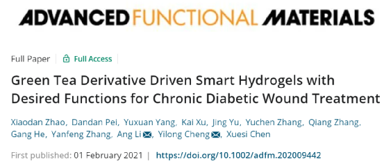
Figure 1 a) Schematic diagram of the preparation procedure of EACPA hydrogel dressing. b) The hydrogel adheres to the authors skin. After peeling, no residue was found. Adequate adhesion can be observed. c) The picture shows the ability of the self-healing EACPA hydrogel to withstand large external forces. d) In the diabetic C57BL/6 mouse model, the mechanism of EACPA hydrogel promoting the chronic wound healing of full-thickness wounds is shown in Figure 2a. The prepared mixture of EA complex and AM aqueous solution (2 m) is at room temperature It is in a sol state and undergoes a sol-gel transition when APS and TEMED are added after purging with nitrogen for 5 minutes. Scanning electron microscopy (SEM) results showed that all EACPA hydrogel samples exhibited an irregular and porous network structure with a pore size of hundreds of microns (Figure 2b). A higher E-A complex concentration results in a slower degradation rate, which is due to the enhanced crosslink density (Figure 2c). Hydrogels formed using dynamic crosslinking agents generally exhibit promising mechanical properties, such as good recyclability and excellent flexibility. As shown in Figure 2d, the spherical shape of the EACPA hydrogel can be restored to its original state immediately without any damage after compression. In addition, the rod-like shape of the EACPA hydrogel can withstand different deformations, such as twisting and bending, thus exhibiting excellent flexibility. As shown in Figure 2e, the tensile test of three hydrogels was further carried out. The authors found that the increase of E-A compound in the system leads to an increase in tensile stress and elongation at break. The hydrogel with the highest E-A composite content (9 mm) showed the highest tensile strength (29.0��6.6 kPa) and the highest elongation at break (661.6��43.4%). The detailed results are summarized in Figure 2f.
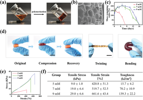
Figure 2 The morphology, degradation curve and mechanical properties of EACPA hydrogels. Materials based on borate bonds will continue to undergo dynamics between reactants (boric acid derivatives and catechol) and products (boronic esters) under field conditions Exchange, so the material usually has excellent self-healing properties. As shown in Figure 3a, when the separated hydrogel surfaces are in contact, the rearrangement of the borate bonds can immediately promote the healing of the hydrogel. For practical applications, the rapid self-repair of the dressing after rupture under normal body movement and local stress can prevent external infections and maintain a sterile environment for wound healing. As shown in Figure 3b, the hydrogel sheet stuck to the authors finger was cut into two parts to mimic the rupture of the dressing. After the two parts are in contact for 30 s, the repaired hydrogel can withstand the bending of the fingers back and forth, showing excellent practicality. Then a rheological study was performed to evaluate the self-healing behavior of EACPA hydrogel (Figure 3c).
Figure 3 Self-healing and tissue adhesion properties of EACPA hydrogel. a) Schematic diagram of the self-healing process of EACPA hydrogel. b) Macroscopic observation of the self-healing effect of EACPA hydrogel adhered to the finger. c) G and G of the EACPA hydrogel dressing using 9 mm E-A complex in five cycles between 1% and 1000% strain. d) The tensile stress-strain curve of the hydrogel after healing at room temperature for 30 s, 3 minutes and 10 minutes. e) Summary of self-healing efficiency of EACPA hydrogel after recovery at room temperature for 30 s, 3 minutes and 10 minutes (n = 5). f) Photograph of tensile test of EACPA hydrogel healed for 10 minutes at room temperature (red circle is the cut part of hydrogel. g) Adhesion to fresh boneless pork slices and image of scalability of EACPA hydrogel . h) Adhesion strength of EACPA hydrogel to pig skin tissue, pig muscle tissue and glass slide The cell biocompatibility of EACPA hydrogel passes (3-(4,5-dimethyl-2-thiazolyl) -2-5,5-Diphenyltetrazolium bromide (MTT) assay and live/dead staining for evaluation. EACPA hydrogels of different quality and cell culture medium were incubated at 37��C for 24 h, and used after leaching In cell culture (L929 cells), first perform MTT analysis to test the metabolic activity of hydrogel-treated cells. As shown in Figure 4a, when the hydrogel concentration reaches 10 mg mL-1, the cell viability is high after 24 hours of incubation At 95%, there is no significant difference between the experimental group and the control group. The biocompatibility of the cells was observed by the live/dead staining method. Live cells and dead cells were stained with green and red fluorescence respectively, and it was found that most of the L929 cells showed normal morphology The cells treated with 10 times of mg mL-1 EACPA hydrogel showed a similar proliferation trend as a control group (tissue culture plate only) (Figure 4b).

Figure 4 Cell compatibility, antioxidant efficiency, bacterial growth inhibition, and EACPA hydrogel transforms the macrophage phenotype from M1 to M2. The results show that the DPPH clearance efficiency is dependent on the concentration of EACPA hydrogel, and the sample is hydrogel The gel has obvious anti-oxidation, and the efficiency of scavenging free radicals is 80%, while the concentration of the hydrogel is greater than 3 mg mL-1 (Figure 4c). As shown in Figure 4d, obvious fluorescence quenching was observed in L929 cells treated with 10 mg mL-1 hydrogel, and the fluorescence intensity was similar to that of the control group and comparable to the Rosup-mediated positive group. The above results prove that EACPA hydrogel has all the ideal properties and can be used as a powerful wound dressing for chronic diabetic wounds. Therefore, the authors evaluated the therapeutic effect of EACPA hydrogel on a full-thickness diabetic wound model. A diabetic mouse model was established in C57BL/6 mice by intraperitoneal injection of streptozotocin (STZ) for 5 consecutive days, because STZ-induced diabetic animal models have been widely used to study diabetic impaired wound healing. Mice whose blood glucose level basically exceeded 300 mg dL-1 (16.65 mmol L-1) within 4 weeks were used to treat EACPA hydrogel dressings. A full-thickness dorsal skin perforation wound with a diameter of 8 mm was inoculated, and then treated with Tegaderm membrane, EACPA hydrogel dressing or without clothes (control group). The details of the treatment are summarized in Figure 5a. Representative pictures of wounds in different groups at predetermined time intervals are shown in Figure 5b. Macroscopically, the authors found that the healing efficiency of EACPA hydrogel was much higher than that of the untreated group. After 18 days of observation, dry sc was still observed in untreated mice. In addition, the quantitative wound area within the experimental time frame was measured based on the wound photos (Figure 5c).

Figure 5 The therapeutic effect of EACPA hydrogel on diabetic chronic wounds. In order to evaluate the healing effect of EACPA hydrogel dressing from the histological point of view, hematoxylin and eosin (H&E) and Eosin (H&E) and eosin were performed on the regenerated skin tissue collected on the 18th day. Massons trichrome staining (MTS) staining. The histological results are consistent with the wound residues in the area observed in the digital image. In the untreated control group, a large number of inflammatory cells encapsulated in the cyst of can be observed, which indicates that inflammation occurs in the early stage of healing (Figure 5d, white dotted circle). Both the EACPA hydrogel and commercial dressing groups maintained a reduced inflammation environment, which was beneficial to wound healing. The granulation tissue formed at the wound bed in the EACPA hydrogel group was also significantly enhanced on the 18th day and was about 120 ��m thick compared with the commercial film dressing group (Figure 5e). In addition, the tissue collected in the EACPA hydrogel group also showed better tissue structure than the control group and the Tegaderm membrane group. In the EACPA hydrogel group, hair follicles, sebaceous glands, sweat ducts, and squamous epithelium were clearly observed (Figure 5d). ). In addition, MTS was performed to detect the deposition of new collagen in the wound healing area (Figure 5f). As we all know, the inflammatory microenvironment is very important in the process of wound healing and tissue regeneration. The inflammatory phase of chronic wounds is severely prolonged, and it cannot even transition to the proliferative phase. Therefore, macrophages mainly exhibit a pro-inflammatory M1 phenotype and produce a series of inflammatory cytokines (ie IL-1��). And interleukin-6 (IL-6) induce tissue destruction and organ dysfunction; the number of M2 phenotype macrophages, which secrete high levels of anti-inflammatory cytokines and repair growth factors (for example, vascular endothelial growth factor (VEGF) , Epidermal Growth Factor (EGF) to reduce inflammation, regulate granulation formation and promote tissue regeneration, greatly reduced. Since these cytokines and growth factors are highly involved in different stages of the wound healing process, in order to further explore the mechanism of EACPA hydrogel dressings to reduce inflammation and promote tissue healing, the tissue around the wound was collected three days after different treatments (Figure 6a) and The content of chemokines in the wound tissue was first detected by ELISA analysis (Figure 6b).

Figure 6 EACPA hydrogel regulates the inflammatory microenvironment to promote the healing of chronic diabetic wounds. a) The wound tissue was harvested on the 3rd day after the different treatments tested below. b) Measure the concentration of chemokines related to inflammation and angiogenesis in wound tissue by ELISA. c) On day 3, immunofluorescence image of IL-1�� (red) and cell nucleus (blue) in skin wound tissue. d) Quantitative analysis of the relative coverage area of IL-1��. e) Immunofluorescence image of macrophages in wound tissue stained with F4/80 (red) and CD163 (green) after treatment. Link to the paper: doi.org/10.1002/adfm.202009442