The superiority and necessity of the application of biomass-derived cellulose nanocrystals (CNCs) in the field of materials has been the goal of constant efforts to explore, especially revealing that the one-dimensional structure, rod-like morphology, and rigid template of cellulose nanocrystals contribute to the high performance of materials. The positive effects of chemistry and functional enhancement. Magnetic resonance imaging (MRI) has become one of the powerful tools for the diagnosis and treatment of tumors by virtue of its advantages such as no radiation damage, high spatial resolution, and three-dimensional visualization. At the same time, longitudinal relaxation (T1) contrast-enhancing contrast agents such as (such as containing Gd) , Mn element contrast agent) proved by clinical practice to effectively improve the accuracy of diagnosis. Studies have found that the morphology of the contrast agent has a significant impact on the relaxation rate that determines its MRI contrast enhancement effect. For example, the one-dimensional rod-shaped manganese-based nanomaterials have a higher specific surface area, which facilitates direct contact between surface paramagnetic Mn2+ and water molecules. Achieve the effect of significantly improving the relaxation rate of the longitudinal contrast agent. However, most manganese-based contrast agents will cause Mn2+ to be continuously released into the patients body during and after imaging, causing cumulative toxicity problems. Therefore, whether biocompatible and highly crystalline CNCs can be used as rigid templates for manganese-based contrast agents by virtue of their one-dimensional structure and rod-like morphology, using surface chemical control methods to construct functional surfaces and effectively combine with Mn2+ to increase The contact rate of Mn2+ with water molecules and the increase of relaxation rate by adjusting the surface Mn2+ density, thereby achieving the purpose of greatly improving the contrast enhancement effect of MRI under the condition of low manganese content. At the same time, only surface-bound Mn2+ is expected to greatly reduce the high toxicity problem caused by the continuous release of non-templated bulk manganese-based nanoparticles on the surface after Mn2+ contrast enhancement and release.
Based on the above design, the team of Professor Huang Jin and Associate Professor Gan Lin of Southwest University cooperated with scientific researchers at home and abroad to utilize the advantages of CNCs "dimension/morphology/template" effect, high chemical activity and biocompatibility, and adopt esterification reaction. The surface is modified with a small molecule chelating agent (diethylenetriaminepentaacetic acid), and then chelated with Mn2+ to prepare an MRI longitudinal relaxation rate enhancement material (Mn2+@DCNCs) with adjustable Mn2+ anchoring density. This research result was published in Celllulose with the title "Enhancing magnetic resonance imaging of bio-based nano-contrast via anchoring manganese on rod-shaped cellulose nanocrystals".
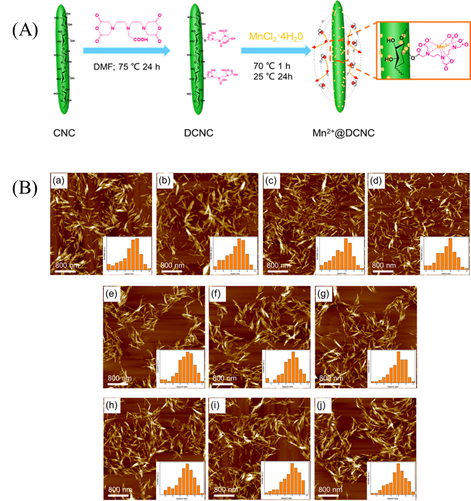
Figure 1. (A) Preparation path of Mn2+@DCNC nanoparticles; (B) (a) CNCs, (b) DCNC-2, (c) DCNC-4, (d) DCNC-8, (e) Mn2+@DCNC -2(1), (f) Mn2+@DCNC-2(2), (g) Mn2+@DCNC-4(1), (h) Mn2+@DCNC-4(2), (i) Mn2+@DCNC-8 (1) and (j) AFM photos of Mn2+@DCNC-8(2) (the illustration is the distribution of aspect ratio)
Due to the high specific surface area of CNCs, the anchoring degree of Mn2+ in this work can be controlled between 0.213% and 0.325%. In addition, the atomic force microscope (AFM) test showed that the rod-like morphology of CNCs was not changed before and after modification, and the aspect ratio was maintained between 17 and 19, which provided the necessary conditions for the increase of longitudinal relaxation rate (Figure 1(B)) ).
Theoretical analysis shows that the longitudinal relaxation (r1) produced by the interaction of Mn2+-H+ can be improved by the rod-shaped morphology, and the degree of improvement depends on the degree of anchoring of Mn2+ on the rod-shaped particles (Figure 2 (a)). The experimental data show that r1 increases linearly with the increase of Mn2+ anchoring degree. It is worth noting that when the Mn2+ content is only 5.9 mmol・g-1, the longitudinal relaxation rate of the nanomaterial can reach 57.71 mM-1・s- 1 (Figure 2 (c)), much higher than conventional magnetic resonance imaging contrast agents. At the same time, because the biocompatible CNCs material is used and only Mn2+ is bound on the surface, the nanomaterial has low toxicity to RAW 264.7 and HUVEC, and the survival rate of the two kinds of cells is higher than 80%. Because this Mn2+-based nanomaterial has high r1 and low cytotoxicity, it has great advantages compared with commercial manganese-based and gadolinium-based MRI contrast agents. Therefore, Mn2+@DCNCs nanomaterials have certain advantages in tumor diagnosis and treatment. potential.
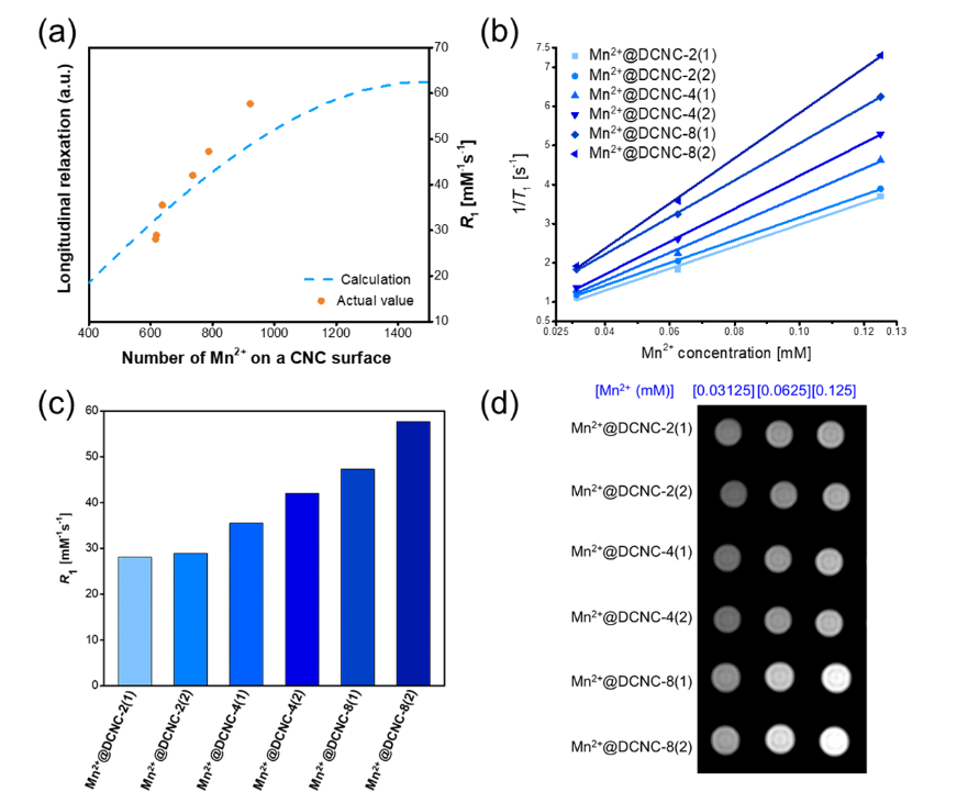
Figure 2 (a) Theoretical and actual data of the influence of Mn2+ anchoring amount on CNC surface on T1; (b) Fitting curve of the relationship between 1/T1 and Mn2+ concentration; (c) Longitudinal relaxation of Mn2+@DCNCs materials with different anchoring amounts; (D) T1-weighted MRI images of Mn2+@DCNCs with different anchors
Chen Yang, a master student in the School of Chemistry and Chemical Engineering of Southwest University, and Dr. Li Xin, Department of Radiology, Union Hospital, Tongji Medical College, Huazhong University of Science and Technology, are the co-first authors. Professor Huang Jin and Associate Professor Gan Lin from the School of Chemistry and Chemical Engineering of Southwest University and Dr. Huang Haitao from the University of Minnesota are the co-corresponding authors. . This achievement has been funded by multiple projects including the National Natural Science Foundation of China (51973175), Chongqing University Innovation Research Group (CXQT19008), Chongqing Talents Program (CQYC201903243) and other projects.
Paper link:
https://doi.org/10.1007/s10570-021-03693-1