[abstract].
The formation of a transparent electrode with seamless contact and optical inquiry at the electrode-brain interface is of potential significance for neuroscience research. Because of its excellent biocompatibility, silk hydrogel can provide an ideal platform for hyaline nerve interface. However, traditional silk hydrogels are too weak to integrate with highly conductive and stretchable electronic devices.
Recently, Zhang Shaomin, a researcher at Zhejiang University and Professor Chen Xiaodong of Nanyang University of Technology, reported a transparent and stretchable hydrogel electrode based on poly (3-ethylenedioxythiophene): polystyrene sulfonate (PEDOT:PSS) and polyethylene glycol fibroin. Polyethylene glycol fibroin with poly (ethylene glycol) Diglycidyl ether (PEGDE) increased Youngs modulus to 1.51 kPa 10.73 MPa and extensibility from traditional silk hydrogel (< 10 kPa) to 400%. Polyethylene glycol wire also helps to form a strong interface with the PEDOT:PSS film, enabling the hydrogel electrode to combine excellent stretching (≈ 260%), stable electrical properties (≈ 4 months) and low thin layer resistance (≈ 160 ±56 Ω) square-1). Finally, the electrodes promoted effective electrical recording and demonstrated the stimulation of barrier-free optical questioning and rat brain imaging. Highly transparent and stretchable hydrogel electrodes provide a practical tool for neuroscience and pave the way for the coordination of tissue-electrode interfaces.The related paper is published in Advanced Materials under the title A Stretchable and Transparent Electrode Based on PEGylated Silk Fibroin for In Vivo Dual-Modal Neural-Vascular Activity Probing.
[guide to the main picture].
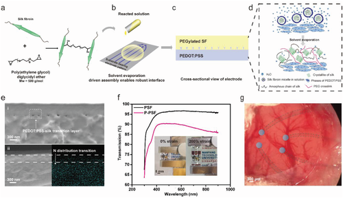
Figure 1.
Design of highly transparent, stretchable and stable hydrogel electrode based on PEDOT:PSS and silk fibroin. A) A schematic diagram showing the formation of cross-linking and ethylene glycol units on SF (PSF) by adding PEGDE. B) the formation of thin film wire electrode. Pour the PSF solution on the PEDOT:PSS and vaporize it. C) Cross-sectional view of the formed electrode, the SF chain (yellow) passes through the interface (P-PSF). D) the formation of P-PSF. The micelle driven by solvent evaporation exposes its chain, thus having more interaction with PSS. E) the cross-sectional SEM image of the transparent P-PSF electrode shows that the interface between PEDOT:PSS and PSF is interconnected (I), and the N distribution at the interface characterized by EDX analysis (ii) shows that N from the wire exists in the PEDOT:PSS region (dotted line). F) the transmission of 300900 nm light through PSF and P-PSF shows that the electrode is highly transparent. Illustration: the optical image of the underwater transparent electrode at 0% (left) and 200% (right) strain proves the stretchability. G) the transparent P-PSF electrode grid was implanted on the surface of rat cortex.
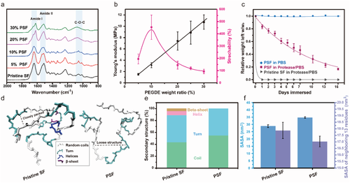
Figure 2.
Molecular characterization, mechanical properties and molecular dynamics simulation of PSF. A) the FTIR spectra of the silk treated by PEGDE with different weight ratios show that the ether part, b) the Youngs modulus (black curve) and extensibility (rose curve) of PSF with different PEGDE wt% under water. C) the biodegradation curves of original SF and 10 wt% PSF in PBS solution with and without protease XIV (1 U mL-1) showed that PSF was stable and biodegradable in PBS. The error bar is the standard deviation of three independent tests. D) representative snapshots of the original SF (left) and PSF (right) models in water show that PSF contains more random coils and turns than the original SF, allowing it to be extended and stretched. E) the quantitative distribution of the secondary structure of original SF and PSF in water confirmed the visual data in (d). F) the solvent accessible space area (SASA) and SASA of the 13 residues at the PEGDE binding site of the original SF and PSF.
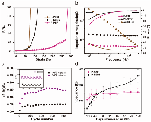
Figure 3.
Electrical properties of P-PSF electrode in wet environment. A) the relative change of PEDOT:PSS resistance on polyethylene glycol SF (P-PSF), PDMS (P-PDMS) and SEBS (P-SEBS) measured underwater. B) Impedance and phase comparison of P-PSF, Pt-SEBS and Au-SEBS in PBS solution at low frequency. C) the cyclic tension of P-PSF at 10% and 30% strain shows that the electrode can last more than 900 cycles. D) the impedance measurements of 100 Hz of P-PSF and P-SEBS incubated in 1 × PBS solution for 4 months showed that P-PSF was highly stable, while P-SEBS showed an eight-fold increase.
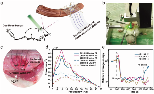
Figure 4.
In situ characterization of neural activity in acute stroke model of photothrombosis in rats. A) schematic diagram of the experimental device. The rose dye was injected into the tail vein of rats and the dye was activated by a laser (green) transmitted by our transparent electrodes on the rat brain to induce acute stroke. B) photographs of rats implanted with P-PSF during photothrombosis. C) the optical image of the P-PSF electrode placed on the rat brain shows that the electrode is transparent and adherent. D) the frequency domain of neural signals recorded in CH1-CH2, CH2-CH3 and CH3-CH4 before and after photothrombosis reflected the local changes of neural activity in core ischemic area (CH2-CH3) and non-core ischemic area (CH1-), in which the attenuation of neural activity in core ischemic area (CH2-CH3) was the greatest. E) the time domain of the neural signal in the frequency range of 0-3 Hz.
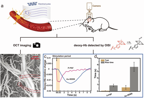
Figure 5.
P-PSF and Au-SEBS electrodes were used to measure the optical imaging and deoxygenated Hb of cerebral vessels after electrical stimulation. A) the schematic diagram shows that the stimulation of neurons leads to the coupling of activated cerebrovascular activity in normal functional rats. B) OCTA images of our P-PSF electrodes on the neurovascular system of the rat brain. C) the representative curve of the relative changes of deoxy-Hb in cerebral vessels after 15 ms current pulse width stimulation at P-PSF and Au-SEBS electrodes. D) Statistical analysis of time of arrival (ToA) and rise time using P-PSF and Au-SEBS electrodes to stimulate cerebral vessels.
[summary].
The team reported a highly transparent, stretchable and stable hydrogel electrode made of PSF and PEDOT:PSS. The electrode is helpful for effective electrical and optical measurement of neural activity in fragile brain tissue. The polyethylene glycol of silk fibroin can form a strong interface with the film PEDOT:PSS. This structure makes our electrodes transparent, stretchable and electronically stable, with high conductivity and patternability. The team applied the electrode to the neurovascular system of rats, and showed that the transparency, low impedance and adaptability of the electrode matched well with optical technology, and could be used to measure the time of neurovascular activity. This kind of electrode which combines electrophysiology and optical function is very ideal. It is hoped that it can be widely used in neuroscience research and clinical applications. In the future, the integration of RF readers will further realize wireless signal transmission and power transmission.
This information is from the Internet for academic exchange only. if there is any infringement, please contact us to delete it immediately.