Metal-organic framework (MOF), as a new type of porous material, is widely used in catalysis, drug delivery, gene therapy, energy storage, etc. It also provides new ideas and directions for bone tissue engineering. 3D printed scaffold is a relatively common artificial bone substitute material. However, the osteogenic performance of 3D printed scaffold loaded with MOF remains to be studied. Recently, the team of Professor Wan Ganbing and Professor Wang Jian of Sichuan University published an article titled "3D printing of metal�Corganic framework incorporated porous scaffolds to promote osteogenic differentiation and bone regeneration" in Nanoscale, and prepared a nanoscale ZIF- 8 (a kind of MOF material) 3D printed porous composite scaffold, the mechanical properties, biocompatibility and bone formation performance of the composite scaffold were studied, and the feasibility of the composite scaffold for bone defect repair was proved.
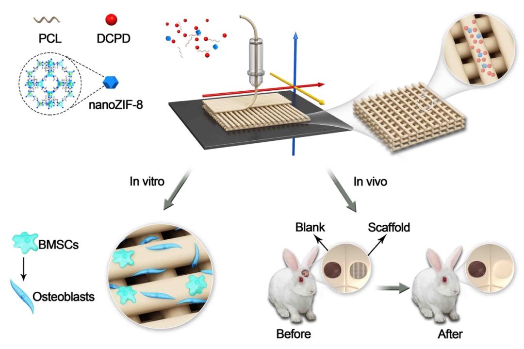
The previous research results of the research group showed that ZIF-8 has excellent in vivo and in vitro osteogenesis, angiogenesis, and antibacterial properties; nano-sized ZIF-8 has lower cytotoxicity and osteogenic properties than micro-sized ZIF-8. In this study, the extrusion 3D printer (EFL-BP-6601) developed by the EFL team was used to prepare a porous composite scaffold loaded with nanoscale ZIF-8, and to explore its bone formation properties in vivo and in vitro. Figure 1 shows that the particle size of the synthesized ZIF-8 reaches the nanometer level; the XRD results of ZIF-8 and the composite scaffold are consistent with their characteristic peaks; the thermogravimetric results show that the ratio of organic matter PCL to calcium phosphate ceramic DCPD in the composite scaffold is about 4:1. Conducive to the stable degradation of the stent; the composite stent also has a slow release effect of Ca and Zn.
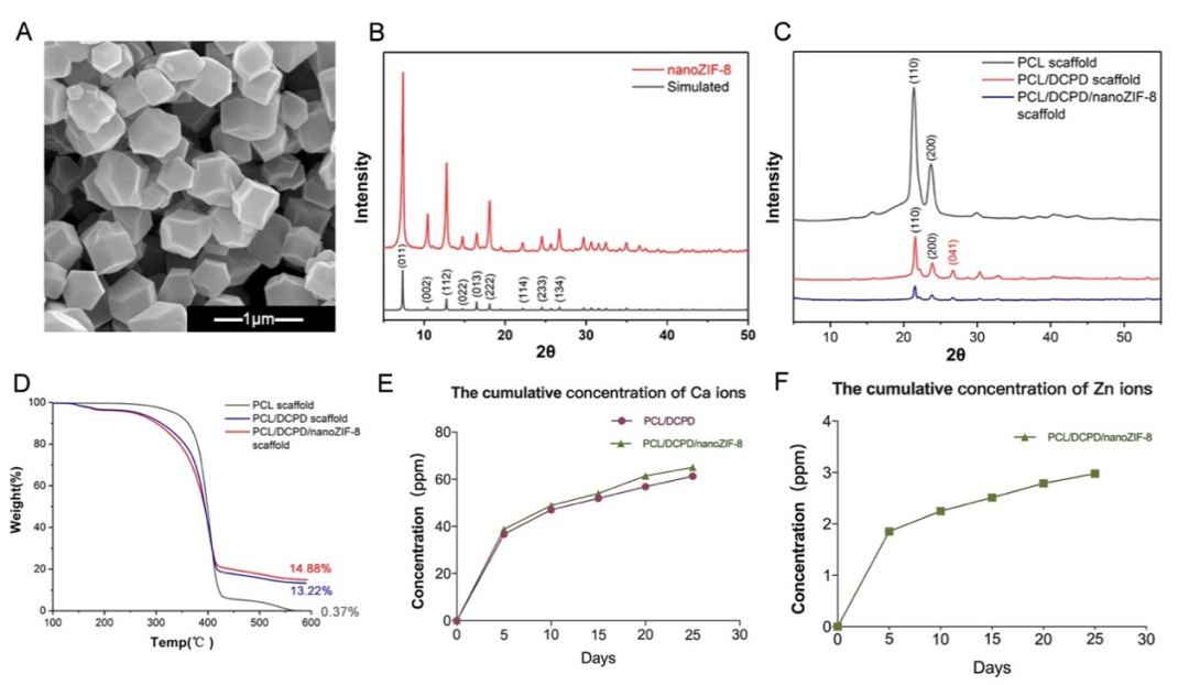
Figure 1 Material characterization of nanoscale ZIF-8 and 3D printed porous scaffold

Figure 2 shows that the three sets of scaffolds have a consistent and uniform structure: a pore size of about 450 ��m and a porosity of 70%. Past studies have shown that it is beneficial to early blood vessel ingrowth and new bone formation; the rough surface of the scaffold is beneficial to cell adhesion. It provides conditions for the later stage of osteogenesis and differentiation; suitable mechanical strength also ensures that the composite scaffold can bear a certain stress and maintain the stability of the environment at the bone defect.
Figure 2 Surface morphology and mechanical properties of 3D printed porous scaffold
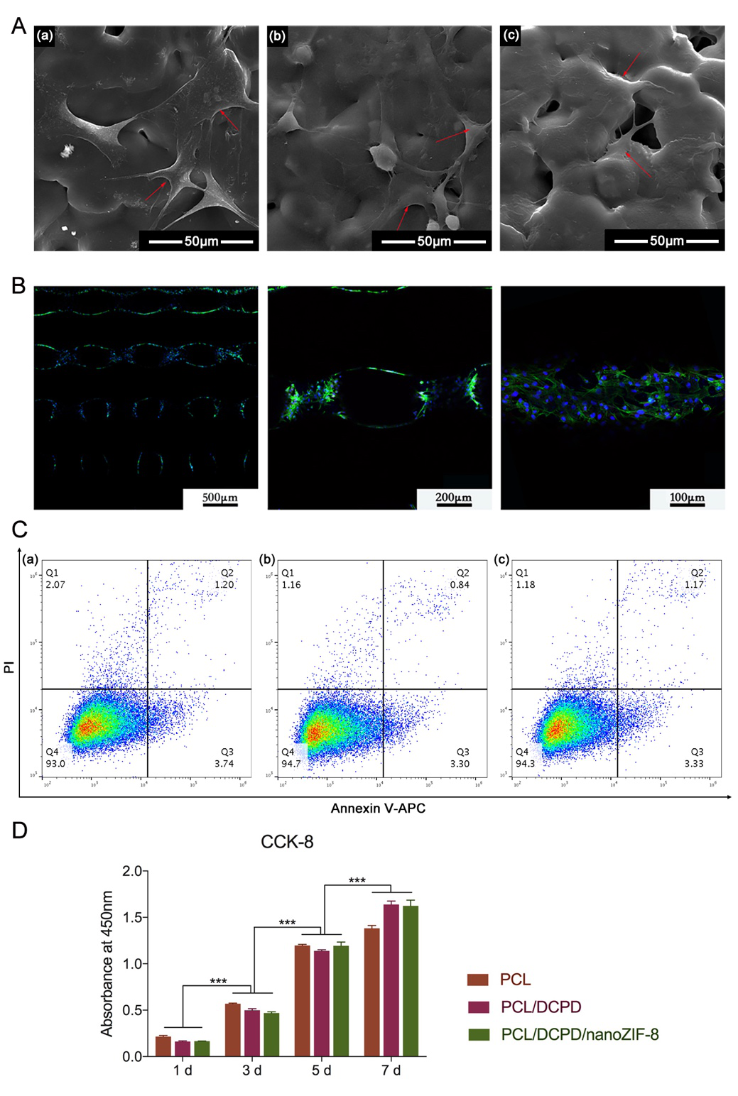
Figure 3 shows that bone marrow mesenchymal stem cells (BMSC) adhere to the surface of the scaffold, fully stretched in a fusiform shape; the 3D scaffold structure has a larger specific surface area, and the cells are distributed on the surface of the scaffold fiber and the inner wall of the pore; the cells on the scaffold surface show The higher proliferation rate and lower apoptosis rate show that it has good biocompatibility.
Figure 3 Biocompatibility of 3D printed stents
The author further studied the osteogenic properties of the composite scaffold in vitro. The results in Figure 4 show that the composite scaffold loaded with nano-scale ZIF-8 significantly up-regulates the expression of bone formation-related genes and proteins of bone marrow mesenchymal stem cells, and promotes the extracellular matrix. Mineralization.
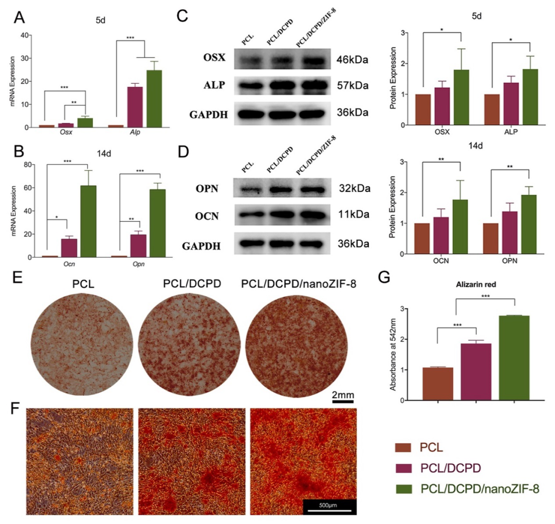
Figure 4 In vitro bone formation performance of 3D printed porous scaffold
The author constructed a rabbit skull defect model, implanted a composite scaffold, and observed the material after 12 weeks. The reconstruction results of Micro-CT (Figure 5) visually show the amount of new bone formation, and there is a large amount of new bone formation in the pores of the composite scaffold loaded with nano-scale ZIF-8. The histological study results (Figure 6) showed that the blank control group had only a small amount of new bone formation around the defect, mostly wrapped in fibrous tissue. Osteoblasts with positive osteocalcin expression can be seen in the pores of the composite scaffold and on the surface of the scaffold. Besides the formation of a large number of new bone on the surface of the scaffold, the formation of vascular structures can be seen.

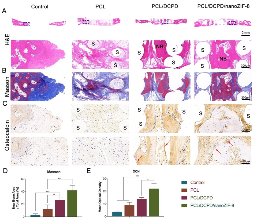
Figure 5, 6 In vivo bone formation performance of 3D printed porous scaffold
In summary, the porous composite scaffold with nano-scale ZIF-8 prepared by extrusion 3D printing technology has suitable mechanical properties, good biocompatibility, and excellent bone formation performance in vivo and in vitro. It is used in the field of bone tissue engineering. Has broad potential.
Paper link:
https://pubs.rsc.org/en/content/articlelanding/2020/nr/d0nr06297a
Information source: EngineeringForLife