已传文件:photo/1793409125.png
 Vaccination is an important approach for cancer immunotherapy, but its limitation lies in the difficulty of efficiently delivering immunogenic antigens to dendritic cells (DCs) to trigger tumor-specific immune responses. In this study, we developed glycosylated lipid nanoparticles (GluLNPs) to deliver tumor antigens and form personalized vaccines in situ, which allows efficient targeting of DCs and stimulates strong anti-tumor immunity, specifically for immunologically cold breast cancer. Through intratumoral injection, GluLNPs can efficiently capture and deliver tumor antigens, including neoantigens released from primary triple-negative breast tumors that are eradicated after mild phototherapy using photosensitizer-loaded nanoparticles (ICGNPs), thereby inducing DC maturation and generating robust anti-tumor immune responses. Moreover, when combined with immune checkpoint blockade therapy, GluLNPs can effectively trigger potent systemic anti-tumor immunity against distant metastatic triple-negative breast tumors. This study proposes a practical strategy for developing effective DC-targeted personalized nanovaccines and in situ vaccination to achieve precise and effective cancer immunotherapy.
 This study was published in Journal of Controlled Release under the title "Dendritic cell-targeted tumor antigen delivery by glucosylated lipid nanoparticles for precision tumor immunotherapy".
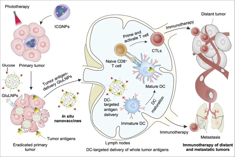
Figure 1
Glycosylated GluLNPs are used for DC-targeted tumor antigen delivery and to regulate anti-cancer immunity against low-immunogenicity solid tumors and their metastases. As an in situ personalized cancer nanovaccine, glycosylated GluLNPs can capture tumor antigens released after ICGNP-mediated phototherapy eliminates the primary tumor, delivering them to DCs. This triggers efficient DC maturation and induces a systemic immune response against low-immunogenicity breast cancer in distant and metastatic tumors.
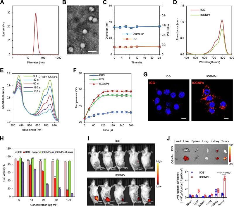
Figure 2.
Characterization of ICGNPs. A, DLS results of ICGNPs. B, Representative TEM image of ICGNPs. Scale bar, 50 nm. C, Stability of ICGNPs under physiological conditions. D, UV‑Vis spectra of free ICG and ICGNPs. E, ¹O₂ production by ICGNPs under NIR laser irradiation. The UV‑Vis spectra of DPBF + ICGNPs (20 μg mL⁻¹ ICG equivalent) after 808 nm NIR laser irradiation (1.5 W cm⁻²) demonstrate ¹O₂ generation, accompanied by a decrease in the DPBF absorbance. F, Photothermal effect of free ICG and ICGNPs (20 μg mL⁻¹ ICG equivalent) under 808 nm NIR laser irradiation (1.5 W cm⁻²). G, Representative CLSM images showing cellular uptake of ICGNPs by 4T1 cancer cells. Scale bar, 10 μm. H, Cell viability of 4T1 cancer cells after phototherapy with ICGNPs. I, In vivo fluorescence imaging of subcutaneous 4T1 tumor‑bearing mice at 24 h after intravenous injection of ICGNPs. J, Ex vivo fluorescence imaging and quantitative fluorescence intensity of tumors and major organs. Data are presented as mean ± S.D. (n = 3 for C, F, H and J). Statistical significance was determined by one‑way ANOVA followed by Tukey’s post‑hoc test; ****P < 0.0001.
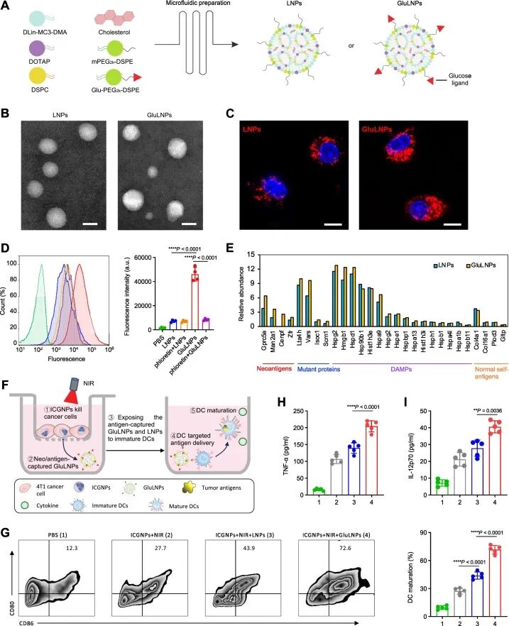 Figure 3.
Preparation and in vitro evaluation of GluLNPs: capturing tumor antigens to form personalized nanovaccines for DC-targeted antigen delivery and activation. A, Schematic illustration of GluLNPs formation. B, Representative TEM images of LNPs and GluLNPs. Scale bar, 50 nm. C, Representative CLSM images showing cellular uptake of LNPs and GluLNPs by BMDCs. Scale bar, 10 μm. D, Representative flow cytometry histograms and calculated fluorescence intensity for cellular uptake by BMDCs. E, Proteomic analysis showing the relative abundance of neoantigens, tumor-mutated proteins, DAMPs and normal self-antigens captured by LNPs and GluLNPs. F, Schematic of the Transwell experimental design: 4T1 breast cancer cells are killed by ICGNP-mediated laser irradiation to release tumor antigens. Subsequently, antigen-captured LNPs and GluLNPs are exposed to DCs for DC-targeted antigen delivery and activation. G, Representative flow cytometry plots and quantitative results of DC maturation induced by antigen-captured LNPs and GluLNPs. H,I, Secretion of TNF-α (H) and IL-12p70 (I) after BMDCs are exposed to antigen-captured LNPs or GluLNPs in the Transwell system. Data are presented as mean ± S.D. (n = 4 for D; n = 5 for G–I). Statistical significance in D and G–I was determined by one-way ANOVA followed by Tukey’s post‑hoc test; *P < 0.05, **P < 0.01, ***P < 0.001, ****P < 0.0001.
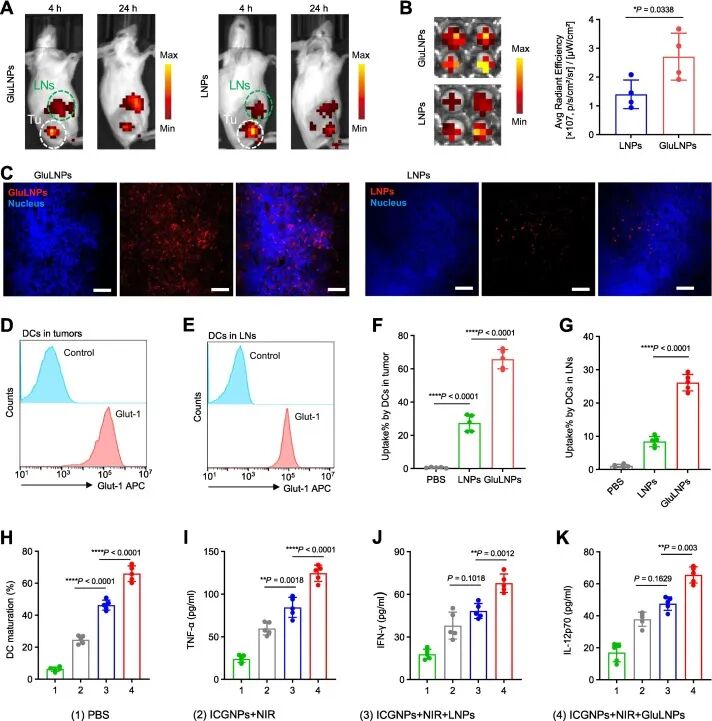
Figure 4.
GluLNPs form in situ nanovaccines after primary tumor eradication by ICGNP-mediated phototherapy, delivering tumor antigens to trigger DC maturation and immune responses. A, IVIS spectra showing the migration of antigen-captured GluLNPs and LNPs from primary tumors to draining lymph nodes after tumor irradiation. B, Images of tumor-draining lymph nodes acquired at 24 h post-treatment reveal greater accumulation of GluLNPs compared with LNPs. C, Fluorescence images of DiR-labelled GluLNPs and LNPs in tumor-draining lymph nodes. Scale bar, 50 μm. D, E, Representative flow cytometry histograms showing Glut-1 expression on DCs in triple-negative breast tumors (D) and draining lymph nodes (E). F, G, Uptake of DiD-labelled GluLNPs and LNPs by DCs recruited to tumors (F) and DCs in inguinal lymph nodes (G). H–K, DC maturation in tumor-draining lymph nodes (H) and serum levels of cytokines TNF-α (I), IFN-γ (J) and IL-12p70 (K) after intratumoral injection of LNPs or GluLNPs into NIR-irradiated 4T1 tumor-bearing mice. Data are presented as mean ± S.D. (n = 4 for B; n = 5 for F–K). Statistical significance was determined by one-way ANOVA followed by Tukey’s post‑hoc test; *P < 0.05, **P < 0.01, ***P < 0.001, ****P < 0.0001.
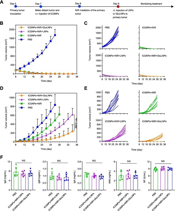
Figure 5
GluLNPs effectively immunotherapy treat distant breast tumors. A, Treatment scheme for mice with primary and distant 4T1 tumors. BALB/c mice were subcutaneously implanted with primary 4T1 tumors on day 0, and with distant tumors on day 7. Treatments included intravenous injection of ICGNPs, NIR irradiation of the primary tumor, and intratumoral injection of LNPs or GluLNPs. B, C, Average (B) and individual (C) primary tumor growth curves. D, E, Average (D) and individual (E) distant tumor growth curves, which were suppressed by the immune therapy effect of GluLNPs. F, Average levels of IgG, IgM, IgA, ANA, and RF in blood after LNPs and GluLNPs treatment. Data in B, D, and F are shown as mean ± S.D. (n=8 for B, D; n=6 for F). Statistical significance in D and F was calculated by one-way ANOVA with Tukey post hoc test; **P < 0.01, NS means not significant.
Summary
The key bottleneck for cancer vaccines lies in the difficulty of efficiently delivering antigens to dendritic cells. We developed glycosylated lipid nanoparticles that, after intratumoral injection, effectively capture tumor antigens—including neoantigens released after phototherapy—and target their delivery to dendritic cells, promoting their maturation and triggering anti-tumor immunity. In a triple-negative breast cancer model, this in situ vaccine combined with immune checkpoint blockade induced a systemic immune response against distant metastases, offering a personalized immunotherapy strategy for poorly immunogenic breast cancer.
This work uses glycosylation modification for dendritic cell targeting and links phototherapy-induced antigen release to the in situ vaccine, eliminating the need for antigen identification and ex vivo loading steps, thereby simplifying the process of preparing personalized vaccines. In the future, other sugar ligands could be screened to suit different DC subsets, or combined with other methods of in situ antigen release. This strategy could be extended to other poorly immunogenic tumors and combined with various immune checkpoint drugs. The long-term safety of the nanoparticles and the feasibility of repeated intratumoral injections still need further evaluation, but this design provides a practical route for the clinical translation of personalized cancer vaccines.
Reference:
DOI: 10.1016/j.jconrel.2026.114671
We specialize in custom nano-medical functional materials. We conduct targeted development to tailor structure, particle size and surface modification for your experiments. Full characterization, iterative optimization and technical support are available for targeted delivery, bioimaging and nanomedicine studies. Send your evaluation plans to us.
Engineer Zhang: 17715390137
Engineer Chen: 18914047343