已传文件:photo/1794019198.png
 Hepatic osteodystrophy (HOD) is a severe metabolic bone disease closely associated with chronic liver disease, and its prevalence is rising rapidly alongside the global surge in metabolic dysfunction‑associated steatohepatitis (MASH). Current clinical interventions remain fragmented and fail to simultaneously address upstream hepatic lipotoxicity and downstream skeletal deterioration. Herein, we engineered copper‑doped bioactive glasses (CuBGs) as a multifunctional ion‑therapeutic nanoplatform. Leveraging their inherent liver tropism, these agents target and reprogram the liver‑bone axis to concurrently ameliorate the pathological manifestations of MASH‑HOD. By delivering local therapeutic ions, CuBGs drive substantial recovery of hepatic metabolism: they improve glucose tolerance, restore mitochondrial oxidative phosphorylation (OXPHOS), thereby effectively suppressing intrahepatic triglyceride accumulation and resolving reactive oxygen species (ROS)‑driven inflammation. Critically, this liver‑rescue mechanism reactivates endocrine crosstalk between the liver and bone by markedly upregulating the secretion of the hepatokine lecithin‑cholesterol acyltransferase (LCAT). Systemic restoration of LCAT directly stimulates osteoblastogenesis and de novo bone formation, efficiently reversing MASH‑induced trabecular bone loss without acting as conventional antiresorptive agents. This dual‑organ regulatory mechanism comprehensively remodels the interconnected multi‑organ microenvironment in MASH‑HOD, with no systemic toxicity observed. Ultimately, this bioactive glass‑mediated copper delivery system establishes a novel, highly scalable ion nanotherapeutic strategy, offering a promising combinatorial therapeutic approach for complicated liver‑bone comorbidities.

This study was published in Bioactive Materials under the title “Bioactive glasses-mediated ion therapy ameliorates hepatic osteodystrophy by reprogramming the liver‑bone axis”.
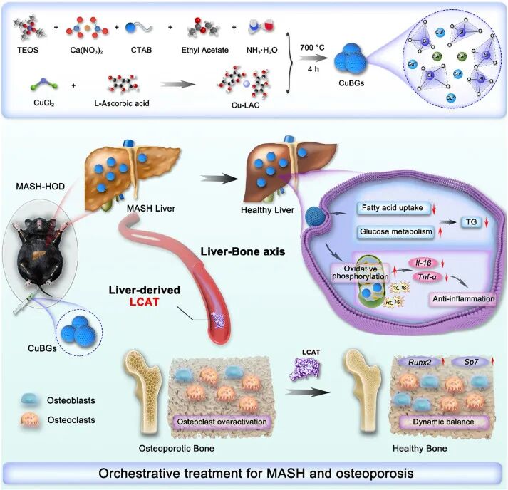 Figure 1
Schematic illustration of the fabrication and therapeutic mechanism of CuBGs.
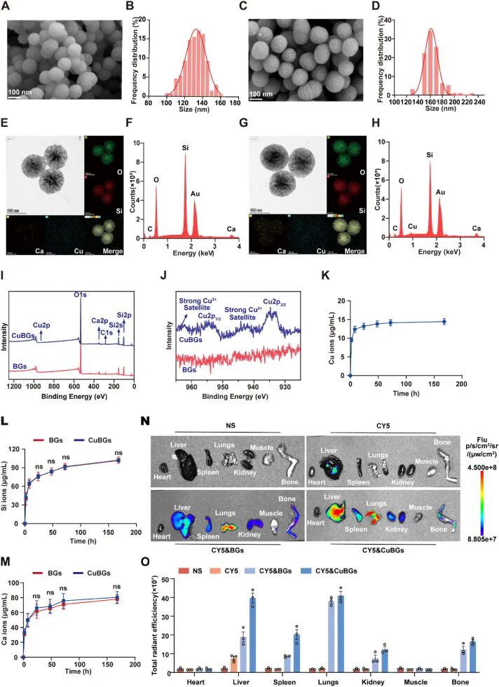
Figure 2
Synthesis, characterization and in vivo biodistribution of CuBGs in mice. (A) SEM image of BGs. Scale bar = 100 nm. (B) Average diameter of porous BGs. (C) SEM image of CuBGs. Scale bar = 100 nm. (D) Average diameter of porous CuBGs. (E and F) TEM image (E) and EDX elemental mapping (F) of BGs. (G and H) TEM image (G) and EDX elemental mapping (H) of CuBGs. (I and J) XPS spectra of MBGNs and Cu‑MBGNs (I), and high‑resolution scans of the Cu 2p3/2 and Cu 2p1/2 peaks (J). (K) Cumulative release profile of Cu ions from CuBGs in serum‑free DMEM over 7 days (n = 3). (L and M) Cumulative release profiles of Si (L) and Ca (M) ions from BGs and CuBGs (n = 3). (N and O) Mice received tail‑vein injection of Cy5‑labelled BGs/CuBGs. (N) Representative IVIS images showing the biodistribution of BGs/CuBGs in major organs at 8 h post‑injection. (O) Quantitative comparison of fluorescence intensity in major organs across different groups.
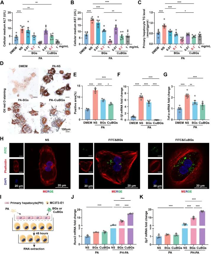
Figure 3
CuBGs ameliorate hepatic injury in MASH and promote osteoblast differentiation in vitro via the liver‑bone axis. (A–C) Primary hepatocytes were cultured in DMEM (control) or stimulated with 0.5 mM palmitic acid to establish an in vitro MASH model. During PA stimulation, cells were simultaneously treated with normal saline, BGs or CuBGs (0.01, 0.1 or 1 mg/mL) for 24 h. (A) ALT levels in the culture medium (n = 6). (B) AST levels in the culture medium (n = 6). (C) Intracellular triglyceride content in primary hepatocytes (n = 6). (D and E) Primary hepatocytes were cultured in DMEM as the normal control or stimulated with 0.5 mM PA to establish an in vitro MASH model. During PA stimulation, cells were concurrently treated with NS, BGs (0.1 mg/mL) or CuBGs (0.1 mg/mL) for 24 h. Representative Oil Red O staining images (D) and quantitative analysis of Oil Red O‑positive area (E). Scale bar = 100 μm, n = 3. (F and G) Relative mRNA expression of Il‑1β (F) and Tnf‑α (G) in primary hepatocytes after the indicated treatments measured by quantitative real‑time PCR. n = 3 biological replicates per group. (H) Primary hepatocytes were incubated with FITC‑labelled BGs or CuBGs for 8 h. Representative images showing intracellular FITC‑labelled BGs or CuBGs (green). Cytoskeleton was stained with rhodamine phalloidin (red). Nuclei were stained with DAPI (blue) (Scale bar = 20 μm). (I) Schematic illustration of the Transwell co‑culture assay. (J and K) mRNA expression of Runx2 and Sp7 in MC3T3‑E1 cells from different groups (n = 3). Values represent mean of 3–5 biological replicates and 1 technical replicate. Data are presented as mean ± SEM, and statistical significance was determined by one‑way analysis of variance. P < 0.05, P < 0.01, P < 0.001.
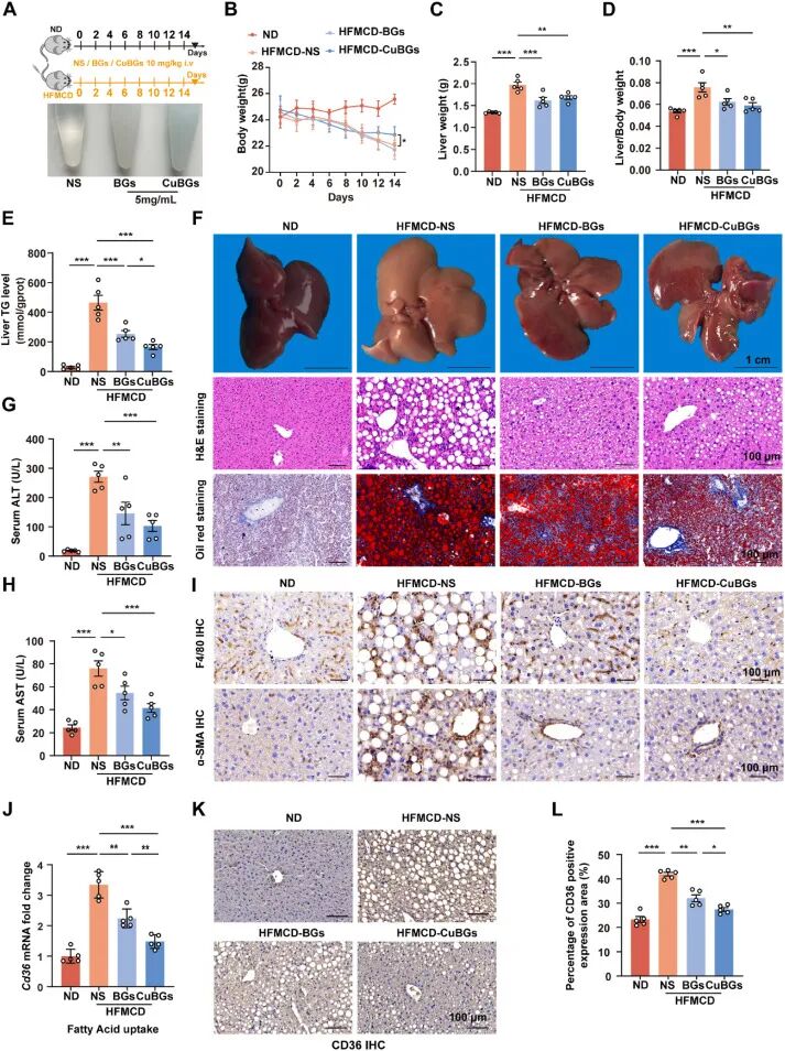
Figure 4
CuBGs ameliorate hepatic injury in the MASH‑HOD mouse model. (A) Schematic illustration of the experimental design (upper) and photographs of injection solutions (lower). Mice were fed a normal diet or a high‑fat methionine‑choline‑deficient diet. During the 2‑week experimental period, HFMCD‑fed mice received tail‑vein injections of normal saline, BGs or CuBGs (particle suspension: 5 mg/mL; 10 mg/kg body weight) every other day, while ND mice received an equal volume (50 μL) of NS on the same schedule. Mice were sacrificed on day 15 after treatment initiation for sample collection. (B) Changes in mouse body weight during the experiment. (C) Liver weight. (D) Liver‑to‑body weight ratio. (E) Hepatic TG levels. (F) Representative gross liver images (scale bar = 1 cm), histological analysis of liver tissues by H&E staining (scale bar = 100 μm) and Oil Red O staining (scale bar = 100 μm). (G and H) Serum ALT (G) and AST (H) levels in different groups. (I) Representative immunohistochemical images of F4/80 staining (upper) and α‑SMA staining (lower) in mouse liver tissues (scale bar = 100 μm). (J) Relative Cd36 mRNA expression in the liver. (K and L) Representative immunohistochemical staining (K) and quantitative expression (L) of CD36 in the liver (scale bar = 100 μm). Data are presented as mean ± SEM (n = 5 mice per group). Statistical significance was determined by one‑way analysis of variance. P < 0.05, P < 0.01, P < 0.001.
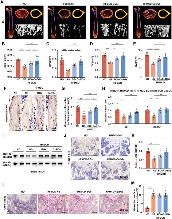 Figure 5
CuBGs treatment prevents bone loss in the MASH‑HOD mouse model. Animal grouping and treatment regimens were identical to those described in Figure 4. (A) Representative 3D μCT images of femoral trabecular bone (scale bar = 400 μm). (B–E) Quantitative analysis of bone mineral density (BMD, B), bone volume (BV, C), tissue volume (TV, D) and bone volume fraction (BV/TV, E). (F and G) Representative immunohistochemical staining for osteocalcin (OCN, F) and quantitative analysis of the number of Ocn‑positive cells per bone surface (G) (scale bar = 10 μm). (H) Relative mRNA expression of osteoblast markers Runx2 and Sp7 determined by quantitative real‑time PCR. (I) Representative western blot of RUNX2 protein levels. (J and K) Representative immunohistochemical staining (J) and quantitative analysis (K) of RUNX2 (scale bar = 400 μm). (L and M) Representative tartrate‑resistant acid phosphatase (TRAP) staining (L) (scale bar = 200 μm) and quantitative analysis of osteoclast surface per bone surface (M). ND: normal diet, NS: normal saline, HFMCD: high‑fat methionine‑choline‑deficient diet. Data are presented as mean ± SEM (n = 5 mice per group). Statistical significance was determined by one‑way analysis of variance. P < 0.05, P < 0.01, P < 0.001.
Summary
Hepatic osteodystrophy is associated with chronic liver disease, and current interventions fail to simultaneously address hepatic lipotoxicity and skeletal deterioration. We designed copper‑doped bioactive glass as an ion‑therapeutic nanoplatform. Leveraging its liver tropism to deliver therapeutic ions, the platform improves glucose tolerance, restores mitochondrial oxidative phosphorylation, and inhibits intrahepatic triglyceride accumulation as well as reactive oxygen species‑driven inflammation. Following hepatic functional recovery, the secretion of the hepatokine LCAT is markedly upregulated, which reactivates endocrine crosstalk between the liver and bone. This directly stimulates osteoblastogenesis and de novo bone formation, reversing trabecular bone loss without conventional antiresorptive agents and inducing no systemic toxicity.
This work targets the liver‑bone axis as an integrated therapeutic unit. A single ion‑based platform achieves coordinated upstream hepatic repair and downstream skeletal protection, overcoming the limitations of fragmented therapies for multi‑organ comorbidities. Future tuning of copper doping ratios or glass degradation rates may accommodate different stages of liver disease and severities of bone loss. As a critical mediator of liver‑bone crosstalk, LCAT can serve as a biomarker for therapeutic monitoring or a direct therapeutic target. This strategy may be extended to other scenarios where metabolic bone disease coincides with liver disorders. Although the long‑term accumulation of copper ions and the safety of bioglass degradation products require further evaluation, this dual‑organ regulatory concept provides a scalable ion‑therapeutic paradigm for complex comorbidities.
Reference
DOI: 10.1016/j.bioactmat.2026.08.028
We specialize in custom nano-medical functional materials. We conduct targeted development to tailor structure, particle size and surface modification for your experiments. Full characterization, iterative optimization and technical support are available for targeted delivery, bioimaging and nanomedicine studies. Send your evaluation plans to us.
Engineer Zhang: 17715390137
Engineer Chen: 18914047343