已传文件:photo/1793586084.png
 Chronic diabetic wounds are notoriously difficult to treat owing to excessive reactive oxygen species (ROS), persistent inflammation, and impaired angiogenesis, which collectively hinder tissue repair. Herein, we report a biomimetic aligned electrospun nanofibrous membrane composed of methacrylated gelatin and o-nitrobenzyl-modified gelatin, functionalized with manganese dioxide (MnO₂) nanozymes for wound microenvironment regulation. Upon hydration and brief ultraviolet irradiation, the membrane transforms into an in-situ nanofibrous hydrogel that conforms to irregular wound beds. Among three MnO₂ loading concentrations, the formulation containing 180 μg/mL MnO₂ was selected as the optimized formulation, as it scavenged intracellular ROS while promoting fibroblast proliferation and migration as well as endothelial angiogenic activity. Mechanistic investigations revealed that the optimized hydrogel stimulated fibroblasts to secrete growth arrest-specific protein 6 (Gas6), which dampened pro-inflammatory macrophage responses and augmented reparative macrophage phenotypes. Gas6 knockdown abrogated these beneficial effects, whereas recombinant Gas6 recapitulated them. Single-cell RNA sequencing uncovered treatment-associated cellular alterations within wounds and identified Gas6-mediated fibroblast–macrophage crosstalk via computational ligand–receptor analysis. In streptozotocin-induced diabetic mice, the optimized hydrogel accelerated wound closure, accompanied by enhanced vascularization, re-epithelialization and collagen remodeling. These findings establish an aligned nanozyme-functionalized nanofibrous hydrogel that integrates redox modulation with Gas6-dependent fibroblast-to-macrophage signaling for diabetic wound regeneration.

This study was published in ACS Nano under the title “Manganese Dioxide Nanozyme-Integrated Aligned Nanofibers Promote Diabetic Wound Repair through Growth Arrest-Specific Protein 6-Mediated Fibroblast-to-Macrophage Communication”.
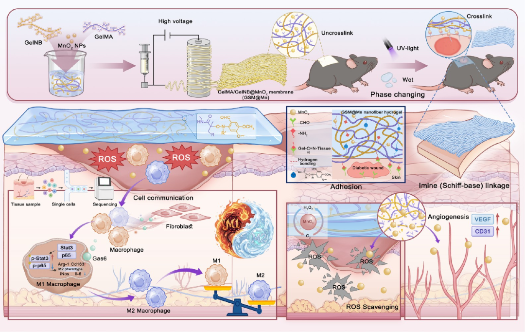
Figure 1
Schematic diagram of the adhesive nanofiber hydrogel driven by nanozymes (GSM@MMn) forming cross-linked electrospun GelMA/GelNB (GSM) films through hydration-induced phase transition and UV-triggered formation. On the optimized intermediate, a MnO2-based NP nanozyme was used at a dose of 180 μg/mL a
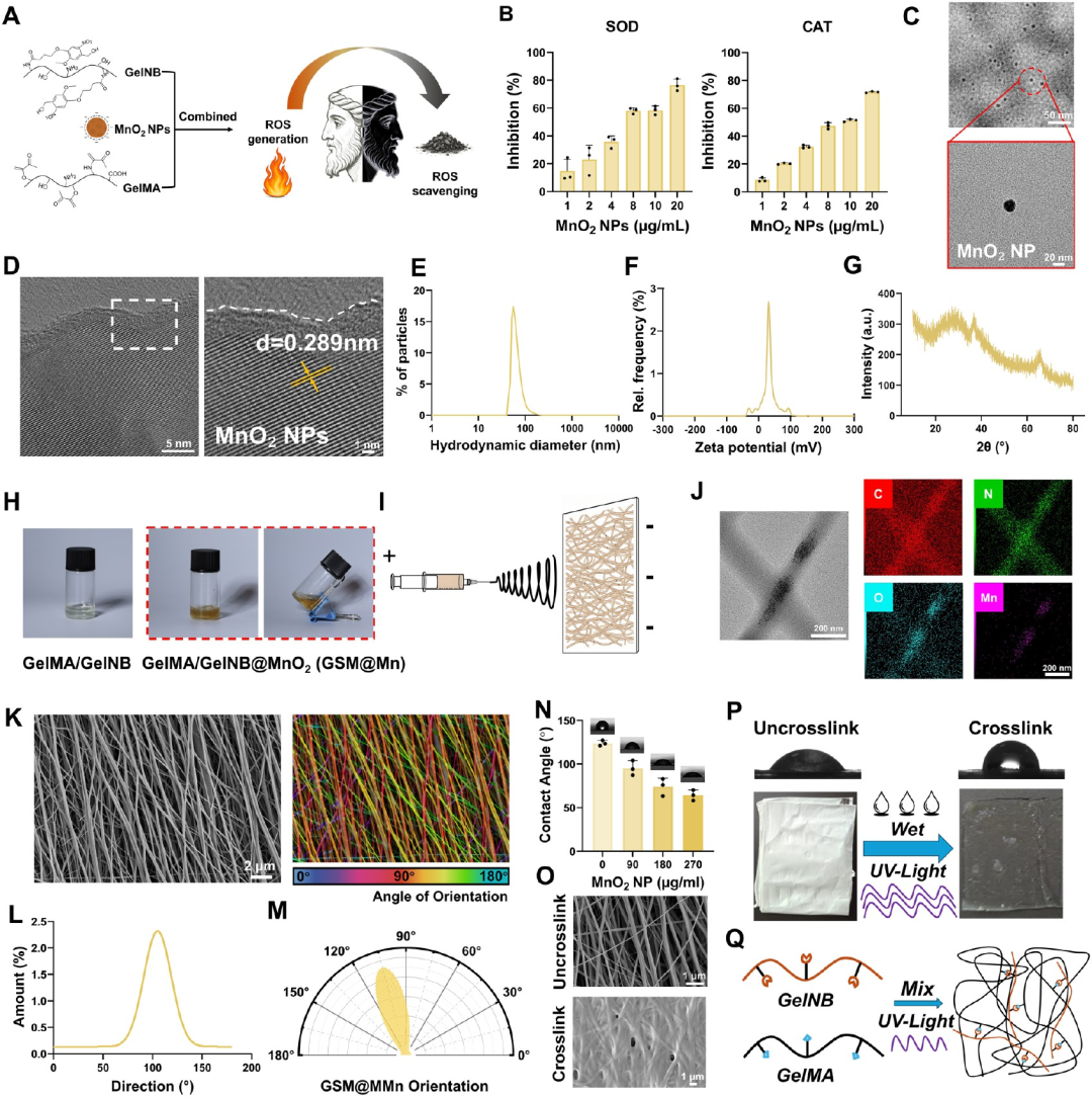
Figure 2
Synthesis and structural design and multi-dimensional characterization of GSM@Mn nanofibrous hydrogels. (A) Schematic illustration of the composition of the GelMA/GelNB (GSM) network and ROS scavenging mechanism based on embedded MnO₂ nanoparticles (MnO₂ NPs) as nanozymes. (B) Quantitative evaluation of SOD-mimetic activity (left) and CAT-mimetic activity (right) of MnO₂ nanozymes with increasing concentrations. (C) Transmission electron microscopy image showing the uniform morphology of as-synthesized MnO₂ nanozymes. (D) High-resolution TEM (HRTEM) image revealing distinct lattice fringes with an interplanar spacing of 0.289 nm. (E–F) Dynamic light scattering (DLS) analysis showing the hydrodynamic diameter distribution (E) and zeta potential distribution (F) of the nanozymes. (G) X-ray diffraction (XRD) pattern of MnO₂ nanozymes. (H) Optical photographs of electrospun precursor solutions of GelMA/GelNB and GelMA/GelNB@MnO₂. (I) Schematic diagram of the electrospinning setup for fabricating aligned nanofibrous membranes. (J) TEM image of a single nanofiber (left) and the corresponding STEM-EDS elemental mapping (right), confirming that Mn is encapsulated within the carbon-, nitrogen-, and oxygen-rich polymer matrix. (K) SEM image of aligned GSM@Mn nanofibers (left) and the corresponding color-coded orientation map (right). Quantitative analysis of fiber alignment is presented in the form of an orientation histogram (L) and a polar plot (M), demonstrating a high degree of alignment. (N) Static water contact angle (WCA) measurements of membranes with varying MnO₂ concentrations, showing a transition from hydrophobic to hydrophilic properties. (O) Comparison of SEM micrographs showing the morphology of dry, uncrosslinked electrospun fibers (top) and the UV-crosslinked hydrogel network after hydration (bottom). (P) Macroscopic photographs demonstrating the transition from a dry fiber mat to a cohesive hydrogel sheet upon wetting and UV irradiation. (Q) Schematic illustration of the proposed photo-triggered transient radical coupled with persistent radical coupling (PTPC) mechanism involved in the formation of the GelMA/GelNB network.
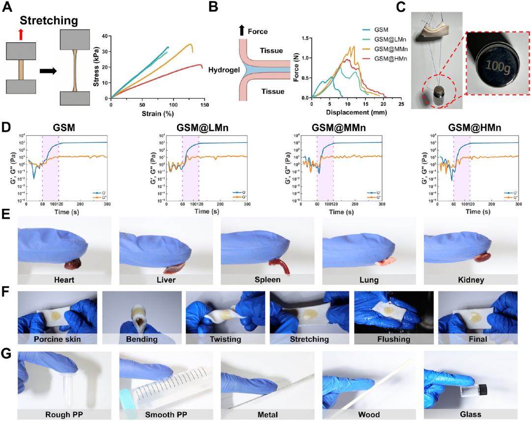
Figure 3
Mechanical, rheological and adhesive characterization of GSM@Mn hydrogels. (A) Schematic setup of uniaxial tensile tests (left) and representative stress–strain curves (right) for GSM, GSM@LMn, GSM@MMn and GSM@HMn hydrogels. (B) Schematic of lap-shear tests using porcine skin (left) and corresponding force–displacement curves (right), showing the tissue adhesion strength of different hydrogel formulations. (C) Digital photograph demonstrating the robust adhesion of the hydrogel, which can stably support a 100 g weight suspended between two bonded pieces of porcine skin. (D) Time-sweep rheological profiles monitoring storage modulus (G′) and loss modulus (G″) during the UV-triggered gelation process of GSM, GSM@LMn, GSM@MMn and GSM@HMn. (E) Photographs showing the adhesion of the hydrogel to various fresh mouse organs, including the heart, liver, spleen, lung and kidney. (F) Demonstration of the adhesion stability of the hydrogel on porcine skin under dynamic mechanical challenges, including bending, twisting, stretching and water rinsing. (G) Evaluation of the versatility of the hydrogel for adhesion to diverse abiotic substrates, including rough and smooth polypropylene (PP), metal, wood and glass.
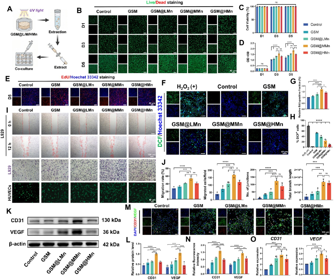
Figure 4
In vitro cytocompatibility, antioxidant capacity, and angiogenic evaluation of GSM@L/M/HMn hydrogel extracts. (A) Schematic illustration of the preparation of hydrogel extracts and the subsequent in vitro cell co-culture setup for detection. (B) Representative live/dead staining images of L929 fibroblasts cultured with different hydrogel extracts for 1, 3 and 5 days (D1, D3 and D5) (live cells: green; dead cells: red). (C–D) Quantitative analysis of cell viability based on live/dead staining (C) and cell proliferation assessment over 5 days using the CCK-8 assay (D). (E) Representative EdU images with Hoechst 33342 nuclear counterstaining (blue) at D5. (F) Representative fluorescence images of intracellular ROS scavenging in L929 fibroblasts challenged with H₂O₂. ROS generation was evaluated by DCFH-DA staining, in which green fluorescence corresponds to ROS accumulation. (G) Quantitative analysis of L929 fibroblast proliferation based on EdU staining. (H) Quantitative analysis of DCF-positive cells for intracellular ROS accumulation. (I) Representative images of the scratch assay for L929 fibroblasts (0 and 12 h), Transwell migration assay of HUVECs (crystal violet staining), and fluorescence images of Matrigel tube-like structures (bottom row). (J) Quantitative analysis of scratch migration rate (%), migrated cell number per field in Transwell, junction number and total branch length (pixels) in the tube formation assay.
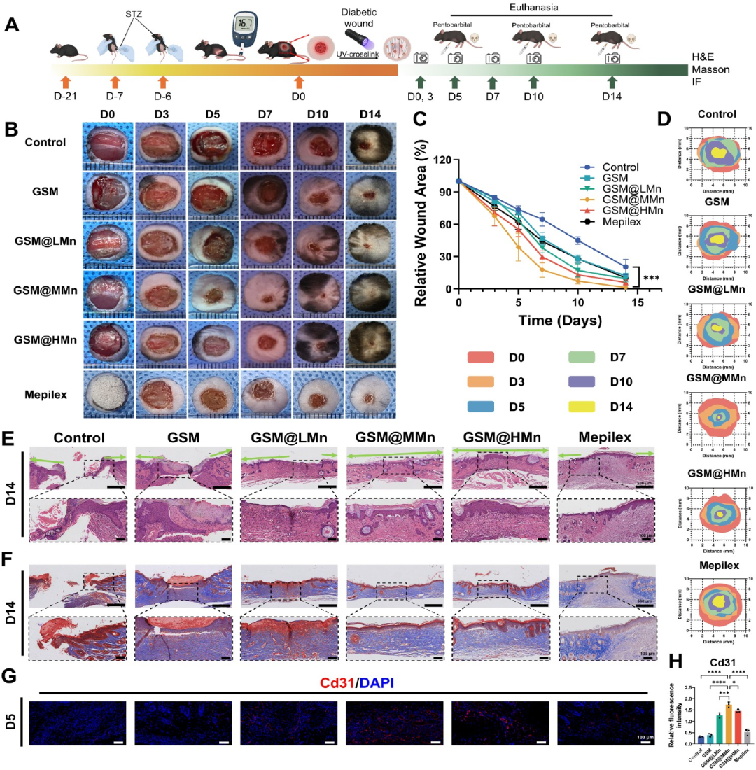
Figure 5
In vivo therapeutic efficacy for diabetic wound healing and histological evaluation of tissue regeneration. (A) Schematic timeline of the animal study, including STZ-induced diabetic model establishment, full-thickness wound creation, wound treatment, sequential photographic examination, euthanasia and tissue harvesting. (B) Representative macroscopic images of the Control, GSM, GSM@LMn, GSM@MMn, GSM@HMn and Mepilex groups on days 0, 3, 5, 7, 10 and 14 post-treatment. Mepilex was used as a comparator commercial foam dressing. (C) Quantification of wound area (%) over time in the six treatment groups, revealing accelerated wound healing with the most prominent effect in the GSM@MMn group (***p < 0.001, GSM@MMn versus Control). (D) Wound tracing maps at sequential time points illustrating the dynamics of wound contraction and closure in each group. (E) Representative H&E staining images of wound tissues at day 14 (upper panels: low magnification; bottom panels: high magnification), showing re-epithelialization and overall tissue architecture. Dotted boxes indicate magnified regions. (F) Masson’s trichrome staining images of day 14 wound sections (upper panels: low magnification; bottom panels: high magnification), showing collagen deposition and remodeling (collagen stained in blue). Dotted boxes indicate magnified regions. (G) IF staining of wound tissues from all six groups at day 5 for CD31 (red) with DAPI nuclear counterstain (blue), for evaluation of early angiogenesis. (H) Quantitative analysis of relative CD31 fluorescence intensity among groups. Data are presented as mean ± standard deviation (n = 3). *p < 0.05, **p < 0.01, ***p < 0.001, and ****p < 0.0001.
mmary
To address the issues of excessive reactive oxygen species (ROS), persistent inflammation and impaired angiogenesis in diabetic wounds, a photocrosslinkable biomimetic aligned nanofiber membrane was designed. This membrane is composed of methacrylated gelatin and o-nitrobenzyl-modified gelatin, loaded with MnO₂ nanozymes. Upon hydration and short-term ultraviolet irradiation, it forms an in-situ nanofiber hydrogel that conforms to irregular wound beds. Three MnO₂ loading concentrations were compared, and 180 μg/mL was selected as the optimal formulation. This concentration attenuates intracellular ROS, and facilitates fibroblast proliferation, migration as well as endothelial angiogenesis. The research workflow first achieved wound conformability and redox microenvironment modulation, followed by formulation screening via cellular functional assays, and finally validated accelerated wound healing, vascularization, re-epithelialization and collagen remodeling in STZ-induced diabetic mice.
The study further attributed the therapeutic effects to the Gas6 signaling pathway. The optimized hydrogel stimulated fibroblasts to secrete Gas6, thereby driving macrophage polarization from the pro-inflammatory phenotype toward the reparative phenotype. Gas6 knockdown abrogated these beneficial effects, while recombinant Gas6 rescued the phenotype. Single-cell RNA sequencing combined with ligand–receptor interaction analysis revealed Gas6-dependent crosstalk between fibroblasts and macrophages. Collectively, this work links nanozyme-mediated redox regulation with intercellular signaling, offering an interpretable and tunable design strategy for diabetic wound therapy. Future investigations will focus on in-depth validation of the Gas6 pathway, material degradation behavior and clinical translation.
Reference:
DOI:10.1021/acsnano.6c14229
We specialize in custom nano-medical functional materials. We conduct targeted development to tailor structure, particle size and surface modification for your experiments. Full characterization, iterative optimization and technical support are available for targeted delivery, bioimaging and nanomedicine studies. Send your evaluation plans to us.
Engineer Zhang: 17715390137
Engineer Chen: 18914047343