已传文件:photo/1793927879.png
 This study was published in Advanced Functional Materials under the title “Preferential Interfacial Engineering of Hydrogels via Interdigitated Nanoparticle Assembly”.
Hydrogels exhibit great potential in diverse fields such as biomedicine, electronics and robotics owing to their intrinsic softness and biocompatibility. Despite these merits, the development of hydrogel-based functional materials and devices is constrained by the limited library of materials that can be seamlessly and robustly integrated with swollen hydrogel matrices. Such interfacial incompatibility substantially hinders their functional expansion and practical deployment. Herein, we report a strategy to facilely integrate hydrogels with a broad range of materials, including polymers, metals and biological tissues, by constructing nanobridges on hydrogel surfaces via the assembly of interdigitated metallic nanoparticles (NPs). This approach relies on ligand-exchange-assisted nanoparticle assembly to induce strong affinity between metallic nanoparticles and the hydrogel matrix. The partially exposed regions of nanoparticles act as nanobridges to mediate chemical or physical bonding with functional components, even hydrophobic ones. Notably, this method enables the effective use of commercial adhesives previously deemed incompatible with hydrogels, broadening their scope for application as biological tissue adhesives. This study establishes a broadly applicable platform for fabricating diverse hydrogel-based systems with versatile interfacial compatibility.
Quick Look at Literature
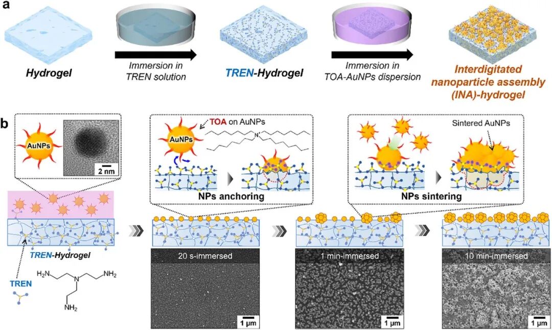
Figure 1
(a) Schematic illustration of the entire process for inducing the formation of interdigitated nanoparticle assemblies on hydrogels via a two-step sequential solution-soaking method. (b) Detailed schematic of the interfacial phenomena at the hydrogel–TOA-AuNP solution interface when the hydrogel incorporated with tris(2-aminoethyl)amine (TREN) is immersed in the trioctylamine-stabilized gold nanoparticle (TOA-AuNP) solution. High-resolution transmission electron microscopy (TEM) images of TOA-AuNPs and top-view scanning electron microscopy (SEM) images obtained at different soaking durations (20 s, 1 min, and 10 min) are provided to clearly demonstrate the time-dependent nanoparticle assembly process and surface evolution.
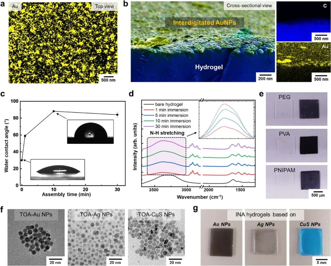
Figure 2
Energy-dispersive X-ray spectroscopy (EDS) analysis of INA hydrogels after 10 min of soaking: (a) Top-view mapping of gold element. (b) Axial elemental distribution profiles; the left panel shows the merged elemental distribution, and the right panel displays separate distributions of carbon (top) and gold (bottom), confirming the uniform incorporation and distribution of gold nanoparticles throughout the hydrogel network. (c) Water contact angle of INA hydrogels as a function of soaking time. (d) Fourier-transform infrared (FT-IR) spectra of pristine hydrogels and INA hydrogels (1–30 min); Inset: magnified view of the 3000–3800 cm⁻¹ region. (e) Photographs of pristine hydrogels and INA-coated PEG, PVA and PNIPAM hydrogels. (f) Transmission electron microscopy (TEM) images of TOA-stabilized gold, silver and copper sulfide nanoparticles. (g) Photographs of INA hydrogels assembled with different nanoparticles.
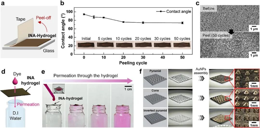
Figure 3
(a) Schematic illustration of the tape-peeling test for INA hydrogels. (b) Variation of water contact angle with the number of peeling cycles. (c) Scanning electron microscopy images of INA hydrogel surfaces before and after repeated peeling (50 cycles). (d) Schematic illustration of the dye penetration test for INA hydrogels. (e) Photographs showing dye permeating through the INA hydrogel and subsequently diffusing into deionized water. (f) Photographs of pyramid-shaped, cone-shaped and inverted-pyramid-shaped hydrogels before and after modification with the INA coating, together with magnified optical images of their surface structures.
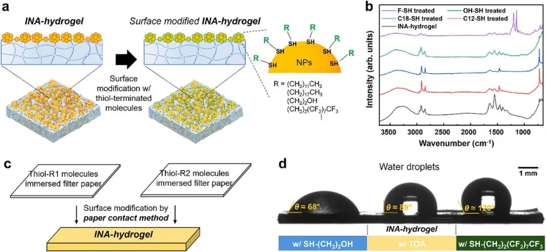
Figure 4
(a) Schematic illustration of interfacial engineering modification of INA hydrogels with diverse small molecules by constructing self-assembled monolayers on the surface of exposed nanoparticle assemblies. (b) Fourier-transform infrared (FT-IR) spectra of INA hydrogels treated with different small-molecule linkers (soaking time: 10 min). (c) Schematic illustration of spatially patterned fabrication of INA hydrogels with distinct surface modifications via the paper-contact method. (d) Photographs of INA hydrogels treated with different small-molecule linkers at separate regions (soaking time: 10 min), demonstrating the patterning capability of this strategy.
Reference Information
Advanced Functional Materials (IF = 19.9)
Pub Date: 2026-08-31
DOI: 10.1002/adfm.78036